 Margen
Did you know? · From the Margen catalogue
Thirty seconds of blue sky and you see your own blood
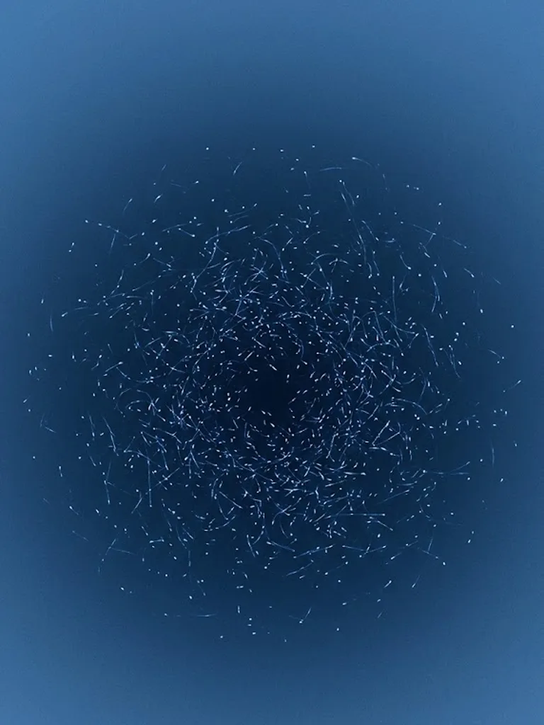
Go outside on a day with a clean sky, lift your face towards a patch of blue with no clouds and no direct sun in it, and hold there for thirty seconds without fixing your eyes on anything. Within a few seconds, tiny bright dots will start crossing, very fast, and they are not in the sky. They are inside your eyes, and they are the trail of your own white blood cells going through the retina.
Watch how they move, because the detail is what matters. They follow short, twisting routes, always the same ones, and they appear only within the fifteen degrees around the point you are looking at. Each one lasts a second or less. And they do something no fly does: they accelerate in pushes, one push per heartbeat.
What you are seeing are gaps, and to see why you have to look at what sits in front of the retina.
In front of the retina's photoreceptors — that is, between the light and the cells that detect it — there is a very fine mesh of capillaries. They are packed with red blood cells, and the haemoglobin inside them absorbs blue light extremely well: the effect peaks at around 430 nanometres, which is exactly the deep blue of a winter sky. Each capillary full of red cells therefore casts a dark line over the receptors behind it. Those lines are there all day and you never see them, because they do not move and the visual system discards whatever does not change.
A white blood cell, a leukocyte, is larger than a red one and does not absorb blue in the same way. When one crosses a capillary in the macula, the centre of the retina, it pushes the red cells aside and opens a transparent window that travels with it. Through that window comes blue light that should not get through, and the receptors underneath light up for an instant.
That explains the three oddities at once. The dots come out bright because what you see is the hole the cell opens as it goes by. They follow fixed, twisting routes because the routes are the capillaries. And they accelerate with your heart because every beat pushes the whole column of blood.
Do not confuse them with floaters. Those are clumps and threads of collagen drifting in the vitreous humour, the transparent gel that fills the eye from inside: they look like strands or grey smudges, they keep sliding after you have already stopped the eye, and they fall slowly. The blue-field ones are smaller than a pinhead, they pass in under a second and they give a tug once per beat. If what you see drags, it is vitreous; if it runs in jumps, it is blood.
If the sky will not co-operate, any uniform and reasonably bright blue field will do: a screen set to blue at full brightness, half a metre from your face, works, and it works better the deeper that blue is. The two conditions are that there must be nothing to look at — no edges, no text, no cloud with a shape — and that you must not fix your gaze on any point, because it is the relaxed eye that lets you see what moves.
In 1980, two researchers turned the trick into a measuring instrument. Charles Riva and Benno Petrig put numbers on it, and years later they patented the method.
The device works like this. The patient is placed in front of a uniform blue field so as to see their own dots. Beside it, a screen generates fake dots, computer-driven, whose speed and quantity can be turned up and down with two knobs. The patient adjusts the knobs until the simulation resembles what they are seeing inside their own eye. When the two match, the simulation's numbers are the patient's numbers, and the patent names the three it returns: how fast their leukocytes travel, how many there are, and how much they accelerate with each beat.
The velocity comes out between half a millimetre and a millimetre per second, which is how far a white blood cell advances along a macular capillary in that time. That speed rises when you breathe air with more carbon dioxide, which dilates the vessels of the retina. And the tug of each beat is counted separately, because it tells you whether that capillary bed is still elastic.
It is a measurement of blood flow in the retina without touching the eye, without injecting anything and without a single needle, and it has been used to study diabetes, glaucoma and the effect of certain drugs on the eye's circulation. It has one serious drawback: the instrument's only sensor is the person. If the patient tires, hesitates or wants to please, the number moves.
Fifty-six years earlier, in 1924, a German ophthalmologist called Richard Scheerer had published the paper that put it in the consulting room. He published it in a German ophthalmology journal, with a title that said exactly what Scheerer wanted to argue: "The entoptic visibility of the movements of the blood in the eye and its clinical significance". In 1924, with that, he was already holding that some little dots you can see by looking at the sky were a clinical measurement. That is why the phenomenon is still also called Scheerer's phenomenon.
But the first person to see his own circulation, and the one who drew it, was working a century earlier and had neither apparatus nor a journal to publish in quickly.
Between 1818 and 1823, before leaving for the chair at Breslau, Jan Evangelista Purkinje, a Czech physician, experimented on his own senses in Prague, with whatever he had to hand. He spun his own body to study vertigo and pressed on his eyes to provoke figures. And one of the things he did was this: to aim a narrow beam of light at the sclera, the white of the eye, instead of at the pupil.
With the light entering from the side, the shadows of the vessels of his own retina stopped falling where they always fall and began falling on neighbouring receptors, which were not used to them. And then Purkinje saw, spread across his own visual field, a tree: the branching drawing of the veins and arteries of his eye, projected onto his sight like an X-ray taken from the inside. The figure is still called the Purkinje tree.
He had nothing to photograph it with: the earliest photograph from life that survives, Niépce's, dates from 1826, and the daguerreotype was not announced until 1839. So he brought the light up to the corner of his own eye, moved the beam slowly so the tree would not go out on him, and drew it by hand.